page forward page back
Table of Contents Index
Dual Chamber Pacing example three
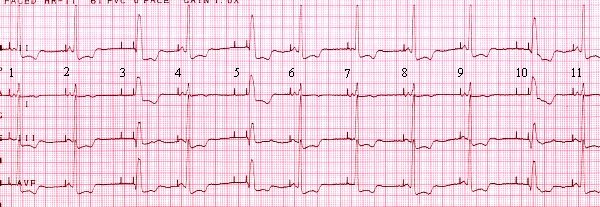
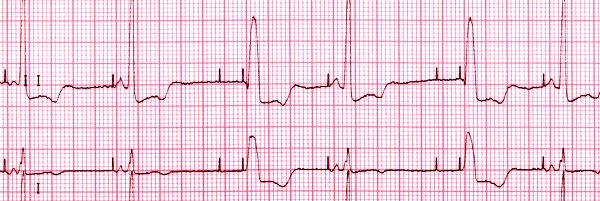
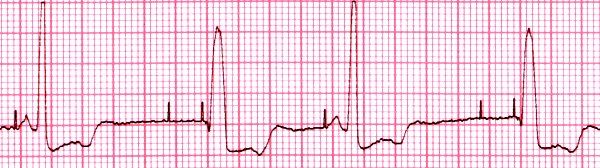
This strip shows some other things a DDD pacer
can do (or fail to do). Beats 1 and 2 are atrially paced, with good capture of the
atria by the pacer, and native conduction from the atria to the ventricles.
Beat 3 shows a pacer impulse stimulating the atria, failure
to capture the atria, a pause as the pacer waits for ventricular
systole (the pacer has been set to wait for 0.21 second, which is just
above the upper limit of normal for a P-R interval), then a ventricular
pacer impulse, which has good capture. Note the difference in QRS
shape: this beat is not coordinated by the specialized conduction system
of the heart. It is wider and slurred, resembling a PVC. Beat
4 is like beats 1 and 2; beat 5 is like beat 3. Beats 6 through 9
are atrially paced, beat 10 is ventricularly paced, and beat 11 is atrially
paced. The pacer is maintaining an adequate heart rate, but the frequent
failure to capture the atria is troubling. Atrial lead parameters
need to be checked. Failure of ventricular capture would be even
more worrisome.