page forward page back
Table of Contents Index
Ventricular Pacing example three
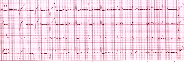
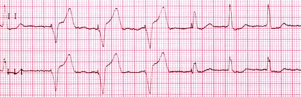
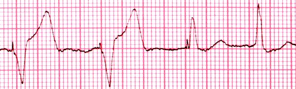
This strip shows native beats, ventricularly paced beats, and fusion
beats.
The underlying rhythm is fine atrial
fibrillation with slow ventricular response.
The first beat is
native: it does not begin with a pacer
spike, it has a QRS width
of about 0.10 second, consistent with a bundle
branch block, a return to
baseline before the T-wave begins, and then a normal appearing T-wave. Beats
2, 3, and 4 are all ventricularly paced beats: they begin with pacer spikes, the
QRS complexes are wide, and the QRS complexes slur into the T-waves, which
are opposite the baseline from the QRS complex. The fifth beat is a fusion beat:
it is produced by two almost simultaneous initiating impulses.
In this case, one
impulse comes from the pacer, and the other is native. The resulting QRS is
different from both the native and the paced QRS complexes.
Beats 6, 7,
and 8 are all native beats, but beats 9 through 12 are fusion beats. Note the
different QRS complexes seen with these fusion beats: depending on the timing
and location of the two originating impulses, many different QRS complexes
can result. The strip ends with a paced beat. The pacer is working in VVI
mode at a rate of 73.