page forward page back
Table of Contents Index
Accelerated Junctional Rhythm example one
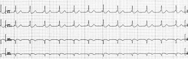

Look at the P-R intervals: they are only 0.08 seconds and the
P-waves crowd
right into the QRS complexes. This P-R interval is too short to represent
normal conduction, and in fact, conduction here
is reversed, or retrograde
(the minimum normal
P-R interval is 0.14 seconds). The pacemaker here is
the junction, and it is pacing the heart at 74 beats a minute. This is much
faster than the junction's normal intrinsic rate, so this is an accelerated
junctional rhythm. The impulse originating in the junction travels into the
ventricles, but simultaneously travels up through the atria in an opposite,
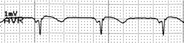
or retrograde, direction. This produces a P-wave that in this case appears
normal in leads II and III, but is inverted in AVR and AVL. Inversion is further
evidence of retrograde conduction:
a reverse flow of depolarization causes
a reversed deflection in the tracing. Except for these inversions
and the
too-short P-R interval, this looks like an atrial rhythm, but those two details
tell the tale.