page forward page back
Table of Contents Index
Premature Atrial Contractions example two
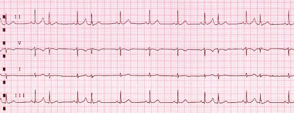
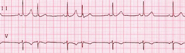
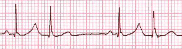
This strip shows several premature
atrial contractions, with interesting features. Notice
that the P-waves that initiate the premature beats are buried in the
T-waves of the preceding beats, changing the shape of these T-waves.
The two voltages are added, so the superimposing of Ps and Ts make a bigger
peak. Also notice that each PAC is followed by the same interval
that separates two normal beats: the rhythm is reset each time.
These two data confirm that the premature beats are atrial beats.
Finally, note that the QRS shapes are not the same. This is best
seen in leads II and III, but can be seen in the other two leads also.
This varying QRS tells us that the specialized
conduction system is functioning differently for the premature beats.
What is happening? Some part of the specialized conduction system
is still refractory when the premature impulse arrives, and the ventricles
depolarize differently. To determine
exactly what the difference is, a 12-lead EKG would be needed.