page forward page
back Table of Contents Index
This atlas is free for you to use, free from advertising, and will stay that way.
If you want to express your appreciation you can send a donation by Paypal to
Alexander Royston Pittman. I will appreciate any amount.
Lead Axis
The axis of an EKG lead is determined by the placement of the lead patches
and which patch is electrically positive or negative. Practically, lead axis is
equivalent to point of view. An inferior lead "looks at" the inferior aspect
of the heart - the apex. A left lead "looks at" the left aspect of the heart
- the
left ventricle. The limb leads are easy to remember: they are numbered
clockwise.
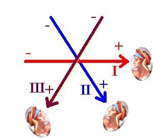
Imagine that the arrows representing these three leads pass through the
heart (seen from the front), and that when examining these leads on an
EKG you are looking down the shaft of the arrow from the positive end.
Lead I is a left lead; leads II and III are inferior leads.
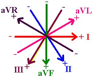
In this diagram, the three augmented limb leads have been added.
They
are easy to remember too: R means right, F means foot, and L means
left.
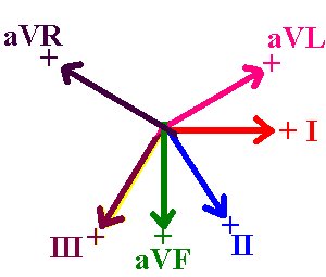
This may make it easier to visualize the lead axes. Only the vector
from the center of the chest to the "point of view" of each lead is shown.
The six V-leads, also called precordial leads, are oriented in a different
plane. They look from the electrode patches on the anterior chest wall
in to the center of the chest. They provide good points of view
to examine
the ventricular septum (V1) and areas in the left ventricle (V2 through V6).